3.2.4 Axons
We model the axons as electrolyte-filled semipermeable membrane tubes with ion channels in their walls. The axons not passively follow the potential’s time course, but they mediate the changes in their internal volume by using an ion pool available in their extracellular volume. The applied potential (including that of the mediated ions) opens the ion channels in the axon’s wall.
In their native mode of operation, the three modes of ion channels define the ’direction of the time’ [62, 127, 26] (the direction of the current that transmits the spike). The layer that the front of the spike creates on the surface (on both sides of the tube) propagates in both directions, but it cannot open the ion channels on the side where the spike arrived from, and the ion channels are still inactivated.
Clamping sets up an artificial working regime for the ion channels: the permanent electric field on the outer surface enables ions to enter the inner volume where formerly no ions (and no potential) existed. The rushed-in ions will flow away from the place of their entrance (recall that the current removes part of the ion layer on the surface), and a slow current toward the membrane can start. Under clamping conditions, the experimenter sets the voltage instead of the transmitted signal and in a static way instead of an autonomous dynamic one.
Initially, the membrane, the clamping point on the axon, and the intracellular and extracellular fluid maintain their resting potential. When an external potential is applied suddenly to some point of the axon, an electric field appears on the outside surface of the axon. The extracellular space with its high ion concentration represents an ”ion cloud” (see also section 2.5.2). When the clamping voltage is switched on, a “fast” current instantly delivers the potential along the outer surface of the axon. However, this is not the case (at least not in the initial moment) on the inner surface. There is no charge present that could change the potential: ’the intracellular concentration at rest is around five orders of magnitude less than that in the extracellular space’ [24]. The physical picture that the clamping potential instantly appears at the end of the axon at the membrane (i.e., if (apparently) they have an infinitely large propagation speed) is valid only if charge carriers exist in the axon.
The persisting clamping voltage gradually triggers the opening of ion channels in its wall along the axon, leading to a continuous inflow through the axon’s wall from the extracellular space into the intracellular space as a ”fast current”; see section 2.8.2. The ions entering the intracellular space remain inside the axon: the cylindrical surface enables only a one-way (inward) traffic for the ions. As discussed in section 11.4 of [24], ”once calcium enters the intracellular cytoplasm it is not free to diffuse”. The ions start to create an ion-rich layer on the internal surface. However, a gradient parallel to the wall exists. The ions experience the electric field (which is present initially only at the clamping point but extends with the passing time) along the axis, speed up, and (after a short while) the ion’s speed becomes constant in time but its value depends on the actual electric field, see Equ. (2.25). The ions will slowly move along the axon with a field-dependent constant velocity in the electric space in a viscous solution. The moving ions deliver charge, so the potential gradually extends along the electrolyte tube (the axon). “In axon fibers, the effective diffusion constant was estimated to be about one-tenth of the diffusion coefficient in aqueous solution” [24]; however, under the effect of the potential gradient, and the mutual repulsion, they form a “slow current” (and that macroscopic current may have a much higher propagation speed). The current and potential are not instant, as we consider in the classic theory of electricity: they propagate with the speed of the ion current.
In this model, we assume that during the time , in the volume , we have a constant ion inflow through the axon’s wall, which increases the charge and concentration already in the volume. The charges in the tube experience the field , and they move with speed inside the tube (see Eq. (2.28)). The ionic fluid with velocity (proportional to ) transfers the ionic charge in the volume to the neighboring element at a distance , and delivers the charge and concentration from the neighboring element at a distance into this element. At the time , the concentration at will result from the inflow at the place (see also the general discussion around Eq. (2.2)). The higher the speed , the more significant the difference between the ”inflow” and the ”present” concentration. The stream inside the axon, a la Minkowski (although in this simple case, a Galilei-transform is sufficient), transforms the distance to time and vice versa. Under the effect of clamping, the current is decreased by the stream proportionally:
| (3.1) |
( is a timing constant of dimension ).
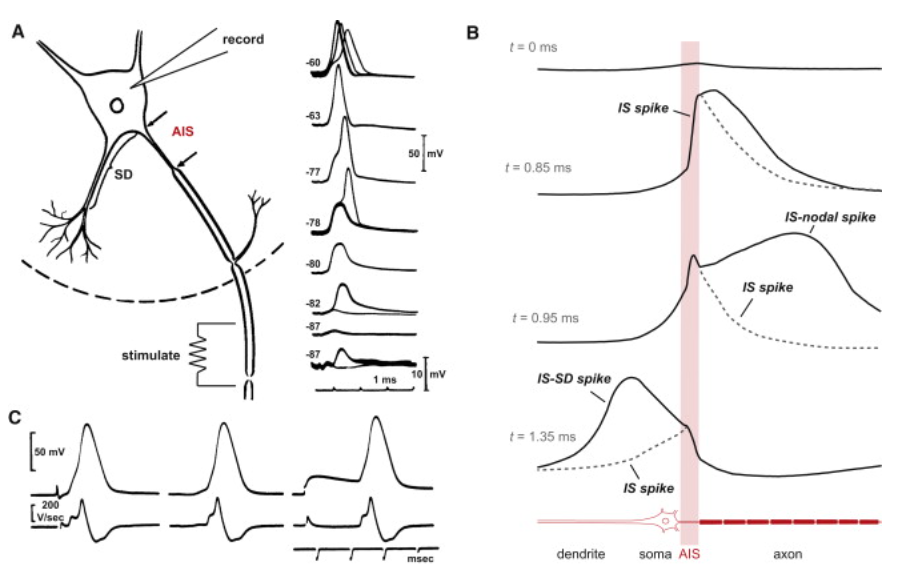
When the stimulation happens inside the axon, and the axon forwards the charge package in the reverse direction [52], towards the AIS. The AIS uses a ”downhill” method of charge forwarding (it is a barrier in both directions), so it can forward the imitated ”AP ” towards the soma. It is a propapation in the reverse direction, since the stimulation arrives from the ”wrong” direction, and the but not a backpropagation. The capacitance of the dendrids explains the shape of the signal.
The AP’s first front already passed to the axon and is forwarded there. It is hard to imagine that the commonly used ”transversal current”, the ion channels, not only synchronize themselves to the length and speed of the spike, but they also sense the direction of the potential gradient.